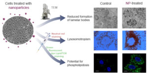
Because of the extremely small size of nanomaterials, they can much more readily gain entry into the human body than larger-sized particles. We are interested in how these nanomaterials behave inside the human body and what influence they have on different human tissues and cells.

We use different cell lines as in vitro models to study the impact of nanomaterials on cell viability and, even more importantly, on the following changes that can be seen already at the subtoxic concentrations: oxidative stress, phospholipidosis, altered enzyme activities, and morphology alterations. We also study cytocompatibility of nanotubular surfaces that can be potentially used for medical application.
Our most recent findings:
- Silica-coated γ-Fe2O3 NPs increase the number of multivesicular bodies and autophagic vacuoles, which are involved in the biogenesis of lamellar bodies in the Type II pulmonary cells A549.
- The genotoxicity of ZnO particles depends on their size: ZnO nanoparticles, but not ZnO microparticles, significantly elevate genotoxicity at the sub-cytotoxic concentrations.
- Different sterilization techniques may significantly influence surface properties of TiO2 nanotubular surfaces and consequently influence their in vitro cytocompatibility.

Methods
- Cell viability assays: MTT assay, neutral red uptake assay, resazurin assay, trypan blue exclusion assay;
- Genotoxicity assays: alkaline comet assay and cytokinesis-block micronucleus assay;
- Scanning and transmission electron microscopy;
- Fluorescence microscopy;
- Spectrofluorometry;
- Flow cytometry;
- Biochemical methods for the measurement of enzyme activity, lipids, phospholipids and carbohydrates;
- Proteomic approaches: SDS-PAGE, 2D-PAGE, western blotting.
Equipment and instrumentation
- Cell culture laboratory equipment;
- Spectrofluorometer (BioTek Cytation, Vermont, USA);
- Inverted phase-contrast microscope (Vert. A1, ZEISS, Germany).
Related content
- Phototoxicity of mesoporous TiO2+Gd microbeads with the potential for cancer diagnosis and treatment (2016, oral presentation); 5th Croatian Congress of Toxicology – CROTOX 2016, Poreč, Croatia
- Cytotoxic and genotoxic effects of ZnO nanoparticles, ZnO microparticles and ZnCl2 on MDCK kidney cell line (2015, presentation); Cutting Edge, Faculty of Chemistry and Chemical Technology, Ljubljana, Slovenia
- Mechanisms of internalization, and sub-cellular localization, of bare and bovine serum albumin-stabilized carbon nanotubes in human alveolar lung epithelial A549 cells in vitro (2015, poster); Final QualityNano Conference, Heraklion, Crete, Greece
- Silica coated magnetic nanoparticles alter the formation of lamellar bodies in pulmonary type II cells in vitro (2015, poster); 1st Slovene Microscopy Symposium, Piran, Slovenia
- Nanoparticles alter the formation of lamellar bodies in pulmonary type II cells in vitro (2015, poster); 6th International Congress of Nanotechnology in Medicine & Biology – BioNanoMed 2015, Graz, Austria